 Approximately eight per cent of men and one-half of one per cent of women in the U.S. have a problem with their color perception.
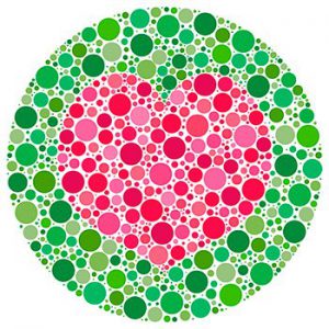
Most colorblind people have lost only part of their color vision. Usually, only one or two types of cones are either absent or not functioning normally. If the dysfunction is in green cones, a person will be deficient picking up green. However, red and blue colors and combinations of red and blue will still be seen.
Causes
In addition to color blindness at birth, color blindness may also occur as the result of traumatic damage to the brain or eye may also develop as a side effect of glaucoma, cataracts, diabetic retinopathy, Alzheimer’s, vitamin A deficiency, allergies to medication, or heart disease.
Color Blindness and the Brain
Unlike color blindness present from birth as the result of genetic defects, “cortical” color blindness can result due to brain damage (such as a stroke). Researchers investigating which part of the brain responds to color discovered that such people see but do not recognize color in the “V4” section of the brain, located at the upper portion of the visual cortex in the back of the head.1
Color-Correcting Lenses
Special vision lenses can help people with color blindness. These lenses, having a custom color spectrum-correcting function can alter the way that color blind people see color.
Custom eyeglasses (based on special color filters called Bragg filters) are effective, but are expensive, bulky, and incompatible with other eyeglasses. Dyed contact lenses are also effective and solve other vision correction issues at the same time.2
In another study gold nanoparticles integrated into contact lens material in three different formations for three types of color blindness, were effective and comfortable.3
Gene Therapy
Partial color blindness
Congenital color blindness is an inherited disorder caused by a single defective or absent gene. About 1 in 12 men lack either the red- or the green-sensitive photoreceptor proteins that are normally present in the color-sensing cells, or cones, of the retina, and so have red–green color blindness. A similar condition affects all male squirrel monkeys, which naturally see the world in just two tones.
In 2009 researchers identified the gene causing the defect and created a gene-carrying virus. They injected the gene-carrying virus into the monkeys’ eyes. In about 20 weeks the monkeys attained full color vision and have shown no harmful side effects. After two years the monkeys still have full color vision with no apparent side effects.4
Gene therapy depending on delivery directly into the retina may have the potential for side effects such as retinal detachment. Researchers are testing a novel intravenously delivered method and find it successful in mice. In any case both methods – direct delivery to the retina and intravenous – appear to effectively reverse color blindness.5
Total color blindness
Total lack of color vision is called “achromatopsia”, a rare genetic congenital condition caused by cone photoreceptor dysfunction. Animal models with achromatopsia have demonstrated partial restoration of color vision. In 2017 gene therapy trials began in three countries. The evidence to date suggests that gene therapy for achromatopsia will need to be applied early in childhood to be effective. 6
Gene therapy for nine patients with achromatopsia was further assessed in a trial published in 2020 in which CNGA3 gene vision disorders were treated. The results were positive, with no substantial safety problems.7
There are a number of ongoing trials for this type of therapy, including trials for related conditions that can cause color blindness such as retinal dystrophy, Leber’s,
Complementary Approach
While alternative approaches cannot cure color blindness, they can support the health of the pigmented cones and vision in general.
Diet. See our diet recommendations.
Nutrients. As cones are found exclusively in the macula of the eye, we recommend taking nutrients that stimulate the macula. These include lutein, bilberry, vinpocetine, gingko biloba, CoQ10, and vitamin A.
Oriental Medicine Approach
TCM refers to color blindness as the “see red as white” condition. It is attributed to prenatal malnutrition. Treatment is to direct, spread and regulate the qi in the channels.
Chinese Herbal Formulas
-
-
- Brighten the Eyes (Ming Mu Di Huang Wan) – Nourishes the liver, enriches the kidneys and improves vision.
- Lycii-Rehmannia (Qi Ju Di Huang Tang) – Tonifies kidney yin, tonifies blood and clears the eyes.
-
Chinese Acupuncture Points
GB 20, LI 3, BL 2, Yi Ming, BL 18, BL 23, KI 3, SP 6, GB 37, Tai Yang
Recommended Supplements
Advanced Eye & Vision Support Formula (whole food) 60 vcaps
Dr. Grossman’s Bilberry/Ginkgo Combination 2oz (60ml).
Dr. Grossman’s Meso Plus Retinal Support and Computer Eye Strain Formula with Astaxanthin 90 vcaps
Vitamin D3 5,000 + K 60 softgels
UBQH 100mg 60 softgels – a well absorbed form of CoQ10
Footnotes
- University of Sydney. (2010). Finding our color center. Retrieved Jul 12 2022 from https://medicalxpress.com/news/2010-11-centre.html. ↩
- Badawy AR, Hassan MU, Elsherif M, Ahmed Z, et al. (2018). Contact Lenses for Color Blindness. Adv Healthc Matar. Jun;7(12):e1800152. ↩
- Salih AE, Elsherif M, Alam F, Yetisen AK, Butt H. (2021). Gold Nanocomposite Contact Lenses for Color Blindness Management. ACS Nano. Mar 23;15(3):4870-4880. ↩
- Dolgin E. (2009). Color blindness corrected by gene therapy. Nature. Sep 16. ↩
- Pavlou M, Schon C, Occelli LM, Rossi A, Meumann N, et al. (2021). Novel AAV capsids for intravitreal gene therapy of photoreceptor disorders. EMBO Mol Med. Apr 9;13(4):e13392. ↩
- Hassall MM, Barnard AR, MacLaren RE. (2017). Gene Therapy for Color Blindness. Yale J Biol Med. Dec 19;90(4):543-551. ↩
- Fishcher MD, Michalakis S, Wilhelm B, Zobor D, et al. (2020). Safety and Vision Outcomes of Subretinal Gene Therapy Targeting Cone Photoreceptors in Achromatopsia: A Nonrandomized Controlled Trial. JAMA Ophthalmol. Jun 1;138(6):643-651. ↩