The retina is a layer of light-sensitive tissue which lines the inside of the eye. When light passes through the lens and hits the retina electrical and chemical agents stimulate nerve impulses and send the message of what is being seen to the brain. The layers of the retina are composed of nerve cells just like the brain.
The early effects of retinal damage may or may not display noticeable changes in vision. If the damage is near the macula, one could notice various visual effects such as general poor vision, distortion of images such as straight lines appearing wavy, blurry spots in one’s central vision, and/or “jack in the box” vision with images appearing and disappearing.
Dim central vision
Distorted central vision
Straight lines that appear wavy
Spots in the central vision that may appear blurry or dark
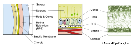
The retina is the most internal layer of the eye. This layer is a very thin, delicate membrane. The term “retina” means “net” or “cobweb” and relates to the appearance of blood vessels within the retina. The retina consists of an outer pigment cell layer and an internal neural layer. In the back of the retina there is a circular depressed area called the optic disc; this is where the optic nerve enters the eye and where its fibers spread out in the neuronal layer of the eye. Because the optic disc contains only nerve fibers and no photoreceptor cells (rods or cones), it is insensitive to light. This is the part of the eye responsible for creating the blind spot that we all notice, most often when we drive.
The outer neural layer which contains nerve cells and blood vessels;
the photoreceptor layer which is a single layer containing the light sensing rods and cones;
a pigmented retinal epithelium (RPE), with the bruch’s membrane separating the RPE from the choroid layer; and
the choroid layer, consisting of connective tissue and very fine capillaries known as choriocapillaries. The tiny capillaries are responsible for carrying nutrients and oxygen to the cellular layers above them.
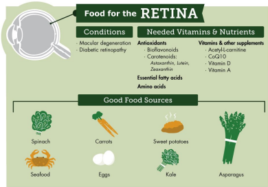
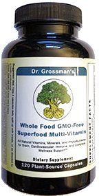
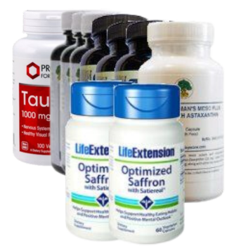
Certain nutrients such as lutein, zeaxanthin, vinpocetine, l-lysine, bilberry, taurine, alpha lipoic acid, lycopene, and a number of vitamins and enzymes and fish oil support the health of these retinal layers.
Also see our juicing recipe to help support retinal health. Juicing is a great way to quickly make nutrients available to your digestive system.