One type of impairment of retinal connective tissue is when a small hole develops in the macula. This impairment takes place where the vitreous adheres to the retina at the macula.
As we age, especially after 50, the vitreous humor which is the gel in the eye, contracts and pulls away from the surface of the retina. This is generally a mild separation without noticeable negative effect. More floaters might result but without significant visual damage.
However, sometimes where the vitreous connective tissue is very firmly attached to the retina surface it can actually pull on the retina eventually forming a small hole in the macula. In addition, with aging the vitreous fluid is less gel-like and more liquid. In that condition it can more easily seep through such a small hole causing a defect or dark spot in central vision which manifests as loss of or distortion of central vision, as well as cause additional pulling on the retina.
Symptoms
Symptoms vary depending on whether the hole is small or extends the full thickness of the macula.
Distorted or wavy vision
Foggy central vision
Difficulty in close work tasks like reading
Blind spot or gray area in central vision
Causes
Very rarely injury to the eye can lead to this condition. Usually, however, these holes apparently develop spontaneously leaving little known method of preventing their development. It is unknown who is at risk.
Conventional treatment
A surgical procedure called vitrectomy is often used to treat holes that go all the way through the macula. The vitreous gel is removed allowing for a variety of repairs, including treatment of macular holes. The gel is replaced with a gas bubble that eventually fills with natural fluids.
Following surgery, patients must usually keep their faces down for two or three weeks. This position allows the bubble to press against the macula and seal the hole. The air bubble itself, however, may take anywhere from 6-8 weeks following surgery to be gradually reabsorbed by the body, and the vitreous cavity is then filled with liquid produced by cells in the front of the eye.
Vitrectomy can lead to complications, most commonly an increase in how fast cataracts develop. Other less common complications include infection, retinal detachment or acute angle-closure glaucoma8 either during surgery or afterward.
Vitrectomy for newly formed (6 months or less) macular holes can result, on average, in about 3 lines on the eye chart improvement. Recovery of vision varies. Some patients achieve only a small amount of vision recovery, while others achieve a more significant improvement.
Inverted internal limiting membrane technique. Another technique involves peeling the internal membrane which surrounds the hole.9 Possible side effects include reduced macular pigment density and impaired linkage to the optic nerve.
Self Help
Although surgery is considered the only treatment, with good nutrition for our vision we may prevent vision problems such as a macular hole. About 50% of foveal detachment macular holes can heal by themselves. Since we consider many eye diseases to reflect the health of the whole body, lifestyle choices and diet can play an important role supporting this healing.
The thickness/density of the macular pigment is associated with incidence of macular holes.1
Research suggests that a number of nutrients including but not limited to zeaxanthin,2, 3, 4 lutein,2, 3, 4, vinpocetine, l-lysine, DHA,4 and specific vitamins and enzymes may help preserve vision.
Low levels of hyaluronic acid, also known as hyaluronan, is a contributing factor in vitreous deterioration, retinal detachment and formation of macular holes.5, 6 Supporting the health of the vitreous and the vitreous body is an important preventative and may assist in preventing or repairing macular holes.
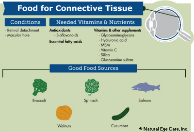
Collagen is a protein and major component of the body from bone to connective tissue. Research has demonstrated its beneficial effect on skin tissue and connective tissue support. Supplementation with collagen may help protect the integrity of the eye’s connective tissue and protect against macular holes. Collagen is abundant in many foods that support good vision such as fish, red and orange vegetables and dark leafy greens.
Some research suggests that daily use of Microcurrent Stimulation (MCS) may help strengthen vision health as well.
Today my husband saw a retinal surgeon due to his macular hole that I spoke with you about 2 weeks ago. Since speaking with you I made fresh carrot juice for him everyday and he followed the product instructions below. The surgeon said the hole is closing and he will not have to have surgery. I attribute this to the juice and supplements. Thanks again for your help and for the discount you gave me.
Sharon C., August, 2017
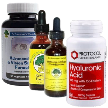
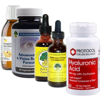
Editor’s Note: The supplements her husband has been taking are the following: Advanced Eye and Vision Support Formula, Retinal Support (wild-crafted herbal formula). Hyaluronic Acid (vegetarian) 100 mg 60 vcaps and Dr. Grossman’s Bilberry/Ginkgo Combination 2oz (60ml)
Our Products
Advanced Eye & Vision Support Formula (whole food) 60 vcaps
1. L. Sauer, S. Peters, et al., Monitoring macular pigment changes in macular holes using fluorescence lifetime imaging ophthalmoscopy, Acta Ophthalmologica, October, 2016. 2. J. M. Stringham, et. al., Macular carotenoid supplementation improves disability glare performance and dynamics of photostress recovery, Eye & Vision, November, 2016. 3. V. Meyer Zu Westrup, et. al, Changes of macular pigment optical density in elderly eyes: a longitudinal analysis from the MARS study, International Journal of Retina and Vitreous, June, 2016. 4. S. Fujimura, K. Ueda, et al., Preliminary analysis of the relationship between serum lutein and zeaxanthin levels and macular pigment optical density, Clinical Ophthalmology, October, 2016. 5. K. Kaprinis, et al, Decreased hyaluronan concentration during primary rhegmatogenous retinal detachment, European Journal of Ophthalmology, November, 2016. 6. B.A. Filas, Q. Zhang, et al, Enzymatic degradation identifies components responsible for the structural properties of the vitreous body, Investigative Ophthalmology & Visual Science, January, 2014. 8. Lee, C.E., Kim, Y.C. (2019). Bilateral Acute Angle-Closure Glaucoma after Macular Hole Surgery. Korean J Ophthalmol, Feb;33(1):101-102. 9. Okonkwo, O.N., Gyasi, M.E., Hassan, A.O., Oderinlo, O. (2018). Inverted Internal Limiting Membrance Technique Maintains Macular Hole Closure in Retinal Detachment Following Macular Hole Repair. Middle East Afr J Ophthalmol, Jul-Dec:25(304):167-169.