 info@naturaleyecare.com


 Home
Home


 Vision
 Vision


 Health
 Health
 Research/Services
 Pets
Pets About/Contact
Cone Rod Dystrophy
Vitamins & supplements Cones Rods Symptoms Causes News
Inherited Eye Problems
Cone rod dystrophy is evidenced by deterioration of photoreceptor cone and rod cells. It is expressed as a number of inherited eye problems, caused by genetic changes in proteins necessary for proper functioning of the photoreceptors. The cones and rods transform light into electric nerve messages that transfer to our brain via our optic nerve. Cones are the photoreceptor cells which allow us to see fine details and color and comprise our central vision. Rods are for low light vision and permit night and peripheral vision. The malfunctioning photoreceptor cells be problematic starting in childhood, or may degrade with time.
 Next:
Nutritional support and
treatment for rod or cone dystrophy.
Next:
Nutritional support and
treatment for rod or cone dystrophy.
Cone dystrophy
Cone dystrophy refers to a number of rare eye disorders where the cone cells suffer degradation. Stationary cone dystrophy symptoms appear early in life and remain more or less stable. Progressive cone dystrophy symptoms tend to get worse over time. Symptom severity and speed of onset can vary greatly from person to person. Some researchers use the term cone dystrophy to refer only to the progressive form of the conditions. The genetic code controls the iodopsin pigment in the cones which helps protect them.
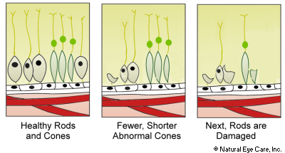
Cone receptors, which are important for color vision and detail, are lost first, followed by rods. The severity of symptoms associated with cone-rod dystrophy vary significantly. Many cases are genetically based, while others are idiopathic (doctors don't know why they develop). Cone-rod dystrophy may first appear in childhood with the failing of some photoreceptor cells, though symptoms may not appear until adulthood. Although complete blindness from cone-rod dystrophy is rare, vision can worsen to 20/200, or less, for those with the progressive form of cone-rod dystrophy.
The stationary form of cone dystrophy is called achromatopsia, meaning vision which lacks color, even though not everyone with this condition is unable to see color. But what does happen is that poorly functioning cone cells cause rod cells to be subjected to too much light. In bright light the vision may appear to be washed out and blurry although the person may be comfortable in darker lighting conditions. Complete blindness does not generally result but patients are often considered legally blind.
Cone dystrophy affects the user's ability to see:
- things that are still
- fine details of things in daylight
- objects in color
- things in fine detail including reading, looking at photographs and recognizing faces
Rod-Cone Dystrophy
Both the cone and rod cells can be affected by genetic mutations - not only the cone cells. Mutations in genetic code undermine the functioning of protective rhodopsin pigment in the rods.
There are two broad groups of CRD:
- Stationary CRD begins to develop during infancy or childhood and symptoms remain stationary for most of life.
- Progressive CRD usually develops a little later, in late childhood or early adulthood. The symptoms progressively worsen to legal blindness, but usually not complete blindness unless the rods also deteriorate.
As rod cells deteriorate then the user has increasing difficulty seeing:
- things that move but only in black and white
- seeing in the dark
- seeing things on the sides of us (peripheral vision)
Symptoms
- gradual loss of night vision
- gradual loss of peripheral vision
- sensitivity to bright light
- vision is best at dusk
- errors in color vision in both red-green and blue-yellow ranges
In young children:
- Parents may notice Nystagmus, which are rapid movements of the eyes back and forth from side to side, and/or
- their child's eyes appearing to slowly wander around not focused upon any object, and/or
- the child touching her eyes with her fingers as though she were poking them.
Parents will often notice these signs by the way the child acts.
Causes
Most causes are genetically based and result from "misprints" in a child's genes. These misprints are typically carried forward from the parents' genes; although sometimes by chance, a misprint occurs in a child's genes when the parents' genes are normal.
There are over 30 types of cone-rod dystrophy caused by mutations in several different genes that can be inherited. Mutations occur in genes that are responsible for making proteins, which are critical in the development, functioning, and health of the photoreceptors. There are several classifications of genetically-based CRD.
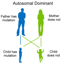
Dominant. Autosomal dominant means that a mutated gene from only one parent is necessary to cause cone-rod dystrophy. For example, the father may have a mutated gene but the mother does not. Each parent gives two genes to children; the mother gives two normal genes; the father gives one normal and one mutated gene and that gene is dominant.
- Gene CRX, known as the cone-rod homeobox gene, is also implicated in Leber’s and retinitis pigmentosa. The CRX gene influences cells in the pineal gland.
- Gene GUCY2D is triggered by abnormal sensitivity to calcium regulation. There has been some investigation into links between this mutation and oxidative stress.
- Gene GCAP1 mutations also cause severe, abnormal-calcium sensitivity and regulation.
- Gene HRG4 is a protein in photoreceptor nerve synapses.
- Loci CRD, the term “loci” refers to the position of genes on chromosomes, and CORD7.
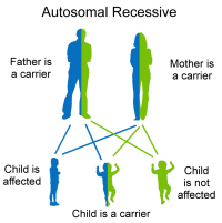
Recessive. Autosomal recessive means that gene mutations must come from both parents. Each parent has one mutated gene and one normal gene. Children may get two mutated (they are affected); one normal and one mutated (they are carriers); or two normal genes and they do not have the mutation.
- Gene ABCA4 causes Stargardt Disease and also 30–60% of autosomal recessive CRDs. Some research suggests that in the absence of healthy ABCA4, tissue is vulnerable to acute oxidative stress. Another researcher mentions the increased oxidative environment in ABCA4 mutations.
- Loci CORD9 and CORD8.
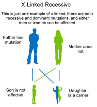
X-linked recessive. Mutations of the X chromosomes can occur in either the mother or the father, and both can pass the mutation to their children. If the mother carries the recessive gene she does not exhibit the mutation. But half of her sons can inherit the mutated x chromosome, and half of her daughters will inherit and be a carrier, but not have the disease. If the father has the mutation, his son is not affected, but his daughter can be a carrier. Men can pass the mutation to their daughters, but not their sons. Usually only men have this form of CRD.
- RPGR mutations cause CORDX1 which is about two-thirds of X-linked retinitis pigmentosa and is also an undetermined percentage of X-linked cone-rod dystrophy.
- An unknown gene (at location xq27) causes CORDX2.
- CACNA1F causes CORDX3. Mutations in this gene also cause night blindness.
- When no cause of CRD can be identified, the case is labeled as idiopathic (cause not known). Also, environmental factors may contribute to causes.
Cone-Rod Dystrophy News
Learn more about this condition in our blog articles on photoreceptor conditions.
Find
Vitamins & Supplements to Support Photoreceptors
Related Conditions
Other eye conditions where the rod and cone photoreceptor cells do not work properly include: Leber's Amaurosis, Retinitis Pigmentosa, Usher Syndrome, and syndromic cone dystrophy (an umbrella term that includes a range of eye diseases, such as Refsum disease, Bardet-Biedl syndrome, NARP syndrome, Batten disease, and spinocerebellar ataxia type 7).

