Stargardt Disease

What it is & symptoms   Causes   Treatment   Complementary Approach   News

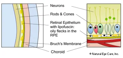
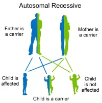
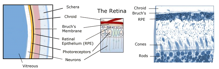
Stargardt disease (fundus flavimaculatus, Stargardt macular dystrophy) is a common type of inherited macular dystrophy affecting young people. It arises from an inherited recessive gene and manifests as a very severe type of macular degeneration beginning in late childhood and resulting in legal blindness.

Next: Nutrients, diet, & lifestyle tips for Stargardt's.

What is Stargardt Disease?

Mutations in the gene ABCA4 (ABCR) underlie Stargardt disease and also have been implicated in cone dystrophy, cone-rod dystrophy, and retinitis pigmentosa. The ABCA4 defect, located on chromosome 1, results in the eyes' inability to transport retinoids (a form of vitamin A) from the photoreceptor cells to the retinal pigment epithelial layer that is responsible for providing nourishment to retinal cells.

Stargardt's is similar to age-related macular degeneration but usually identified before age 20. The first symptom is decreased vision.

The symptoms of Stargardt's are similar to those of age-related macular degeneration (ARMD). It is diagnosed in 1 in 20,000 children over 6, usually identified before age 20. The first symptom is decreased vision.

  • Poor vision in dim light.
  • Difficulty in close vision, such as reading, although general vision may seem fine.
  • Loss of vision sharpness, poor color vision and small blind spots.
  • Blurriness and distorted vision
  • Difficulty adapting to the dark after sunlight exposure, and/or light sensitivity.
  • In more severe stages of vision loss, patients may experience phantom vision or visual hallucinations. These episodes are not usually related to underlying psychiatric problems, but rather are normal attempt by the brain to make sense of impaired sensory information. The brain may embellish the image making it very real just as it does in our dreams.

Early Stage

Stargardt disease results in progressive central vision loss, first reported as difficulty in reading and seeing in low light situations, although vision is generally good. These problems are generally noticed between ages 6 to 12, though the symptoms may also arise as an adult.

Disease Progression

Stargardt diagram With further progression, oily deposits begin to accumulate in the layer of tissue comprising the retinal pigment layer of cells (RPE) which is the layer behind the macula. The RPE is a layer lying between the retina and the choroid, providing nourishment to photoreceptor cells. These deposits, called "lipofuscin", look like yellowish-tinted specks. The accumulation of lipofuscin eventually causes wasting away of the macula and the RPE. How fast and to what degree vision is lost varies, especially in children. It can begin with 20/40 vision and rapidly decrease to 20/200 which is the level for legal blindness. Color vision may also be impaired in advanced Stargardt's. By age 50, about half of all of patients assessed in clinical trials had visual acuities of 20/200 to 20/400.

Causes

Stargardt syndrome

Stargardt disease nearly always arises from an inherited recessive gene. This means that both parents carry the gene. Each of their children has a 25% chance of inheriting a gene from each parent. Parents might not themselves have Stargardt's since they might have only one gene and two are required to cause Stargardt's.

ABCA4 Gene Mutation

In 1997, researchers identified the specific gene tied to Stargardt's. The ABCA4 gene mutation is one of 19 known mutations of the ABCR gene. ABCR is a large retinal protein whose code is regulated by the ABCA4 gene. It controls the production and behavior of the protein responsible for energy transport to and from photoreceptor cells in the retina.

The mutations result in a non-functioning protein that is unable perform its transport function. The dysfunctional ABCR protein allows fatty deposits to accumulate in the retina's RPE layer. The RPE layer gradually becomes damaged causing central vision loss, often first noticed between ages 6 and 12. As the effects of the mutation continue the RPE layer begins to collect oily, granular deposits, called "lipofuscin" which impinge on the healthy structure of the retina. They look like yellowish-tinted specks and form clumps called "vitamin A dimmers."

Stargardt's News

Want to learn more? See our blog for news on Stargardt's.

Stargardt Vitamins Find Vitamins & Supplements to support the retina.