
Vitamins/supplements for retinal support
Best’s disease, also known as Best’s vitelliform macular dystrophy, is a hereditary (usually) form of progressive macular dystrophy.
- Pre vitelliform: Initially a recording of eye movements and eye position identifies abnormal electrical potential. Eyes will be tested resting or moving in dark and light conditions.
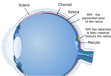
- Vitelliform: Usually occurs between 10-25 years of age. Typical yellow spots, sometimes accompanied by material leaking into a space by the retina can be observed; an observation called “egg-yolk” lesion.
- Pseudohypopyon: When part of the lesion becomes absorbed, this is identified as stage three. Even at this stage there may be little effect on vision.
- Vitelliruptive: At this stage, when the “egg-yolk” breaks up in a process referred to as “scrambled-egg”, sight will probably be affected.
- Atrophic: The fifth and final stage is when the condition causes the most severe sight loss.
- Plus a Complication: Choroidal neovascularization results in about 1 in 5 patients.
Stages
Symptoms
Gradual loss of central vision.Causes
Best’s vitelliform macular dystrophy is a condition that tends to run genetically in families due to a mutation in the VMD2 gene. The mutation can be transmitted by only one parent. Without any family history of Best’s disease, about 25% of Best’s patients will also have this genetic mutation. Researchers have found over 40 mutation variations in this gene. The researchers also concluded that a few patients who are diagnosed as having macular degeneration (AMD) actually have late onset Best’s.1
The VMD2 gene encodes bestrophin, a protein form which is found in a wide variety of organisms where it is thought move chloride atoms around in the retinal cells. This process regulates pH levels, movement of solutes among cells, cell growth and cell movement. Mutations in the VMD2 gene, causing malfunctioning of bestrophin permits fatty yellow pigments to build up in the cells between the macula
The related vitelliform macular dystrophy is caused by mutations of either the same VMD2 gene or the RDS.
Conventional Treatment
There has been no conventional treatment, although the literature has a reference to one patient injected with bevacizumab.2 A 2015 report of photodynamic therapy shows some promise. Photodynamic therapy (PDT) involves using a light-sensitive drug, which, when exposed to specific light wavelengths create a oxygen type that kills adjacent cells. The researchers evaluated 30 patients, three of whom had received PDT treatment. There was an average 77 month followup during which there was stable vision functioning followed by slowly improving vision functioning for several years.3 This study substantiated a 2003 report involving a single patient.
Self Help
Lifestyle choices and diet can play a major factor in helping manage Best’s disease. See our homeopathic products and recommendations for supporting the macula in the sidebar.
Certain nutrients such as lutein, zeaxanthin, vinpocetine, l-lysine, a number of vitamins & enzymes, and fish oil may help slow down Best’s Disease and preserve vision.
Daily juicing of vegetables and fruits (preferably organic). Our suggested retinal support recipe is a combination of some the following: ginger, garlic, leeks, parsley, beets, cabbage, carrots, celery, spinach, apples, grapes, raspberries, lemon, chlorophyll, wheat grasses (not too much fruit). See more juicing information.
Best's Disease News
Want to learn more? See our blog news on Best’s disease.

Research
Although the underlying physiological cause may be unique, there may be similarities in terms of nutritional, diet and lifestyle recommendations made by Dr. Grossman for eye conditions that result in similar vision symptoms to those of Best’s Disease.
Footnotes
1. Andrew J. Lotery, et al, Allelic Variation in the VMD2 Gene in Best Disease and Age-Related Macular Degeneration, Biochemistry and Molecular Biology, May, 2000.
2. C. Celea, et al, Evolution of Choroidal Neovascular Membrane in Best Disease after Single Intravitreal Bevacizumab. Case Report, Maedica, March, 2015.
3. A. Sodi, et al, Long-Term Results of Photodynamic Therapy for Choroidal Neovascularization in Pediatric Patients with Best Vitelliform Macular Dystrophy, Ophthalmic Genetics, June, 2015.