 info@naturaleyecare.com


 Home
Home


 Vision
 Vision


 Health
 Health
 Research/Services
 Pets
Pets About/Contact
The Retina
How it works
Retinal layers
Structure
Types of damage
Photoreceptors
Connective
Hole /
Wrinkle in the macula
Sugar balance
Vitamin A
The retina is structured of several layers of nerve cells, photoreceptors, pigmentation and blood vessels which line the inside of the eyeball. The retina spreads around the inside of the entire back of the eye. The central portion of the retina is called the macula, which is responsible for central vision.
How light is converted to nerve impulses
Light passing through the flexible lens, striking the retina and being converted to nerve impulses involves an interesting process. Light has to pass through layers of nerve cells, photoreceptors, pigmentation cells, and blood capillaries before it reaches the retina. Muller cells are living optic fibers that support the nerve cells. They lie outside the retina in long cylindrical tubes that transmit light to the retina.1
The photoreceptors (rods and cones) in the retina cones absorb photons of light, changing shape as they do so. The change in shape initiates chemical and electrical impulses which in turn stimulate nerve impulses sending the message of what is being seen through the optic nerve to the brain.
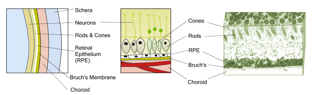
Retinal layers
The retina's2 layers provide for the transmission of light from outside to the brain. A variety of retina conditions arise from malformations in the different layers.
- The outer layer contains nerve cells called ganglions contain axons that transmit optic messages to the brain. This layer of nerve cells and the main optic nerve they connect to are actually brain tissue and are considered part of the central nervous system.
- The photoreceptor layer contains light sensing rods and cones. These photoreceptors are large molecule chemicals that change size when they are energized. This energization is a greater than normal state of energy, and when it 'relaxes' it passes that energy on to nerve endings. In the rods (for dim light and black/white vision), these molecules are called rhodopsin, and in the cones (for color vision), they are known as iodpisin.
- The next pigmented layer is called retinal epithelium or RPE. These cells are a single layer of hexagonal cells of pigment bits tightly packed together. The important pigment of these cells is what protects the retina from the damaging effects of sunlight. The body builds up omega-3 fatty acids in the RPE to maintain the photoreceptor structure, and glucose to provide energy to the retina and other needed components to the choroid and retina. In addition, this thin layer helps drain excess fluid, maintain pH balance and get rid of damaged or dead old, outer photoreceptor cells.
- The Bruch's membrane is a barrier and filter between the RPE and the choroid layer, separating blood from neurons. It includes connective tissue and tiny choriocapillaries that carry nutrients and oxygen to the cells. It is an elastic layer that gets thicker with age, limiting the capacity of capillaries to deliver nourishment and oxygen to the other parts of the retina. The photoreceptor cells require a lot of oxygen for energy and need a constant supply of oxygen-carrying blood, but the neurons require space between the neurons/axons without intrusion of fluid or other wastes. Bruch's membrane supplies this need.
- The choroid layer contains connective tissue with many blood vessels. It delivers nourishment and oxygen to the outer layers of the retina. It consists of Haller's layer, with larger blood vessels, Sattler's layer, with medium sized veins and arteries, and choriocapillaries, a layer of very fine capillaries. Then the Bruch's membrane separates this section from the RPE and the inner portions of the retina.
Retina damage
Retina damage can take a variety of forms:
- General damage to the retina
- Photo-receptor damage
- Connective tissue damage
- Connective tissue (hole)
- Damage to sugar imbalances
Footnotes & Sources:
1. Franze, et al. 2007. Muller cells are living optical fibers in the vertebrate retina. PNAS 104: 8287-8292.
2. Wikipedia sections on retina, retinal epithelium, photoreceptors, bruch's, choroid, etc.,
How it works
Retinal layers
Structure
Types of damage
Photoreceptors
Connective
Hole /
Wrinkle in the macula
Sugar balance
Vitamin A

